
Fertomid
"Fertomid 50mg on-line, breast cancer types".
By: K. Dimitar, M.S., Ph.D.
Clinical Director, Wayne State University School of Medicine
Presented here is a simple procedure for the preparation and study of the genital plate of Drosophila species pregnancy 21 weeks order fertomid 50mg free shipping. Objectives: Dissection and study of Periphallic organ/Genital plate of Drosophila Materials Required: A menopause joint pain relief buy fertomid 50 mg cheap. Fly Stocks: Stocks of different species of Drosophila or freshly collected from the wild breast cancer 900 position purchase fertomid cheap. Select and take a few male flies on a clean plain slide in a drop of saline solution pregnancy 7 weeks purchase 50 mg fertomid with amex. Using fine needles cut and separate out, under a stereo-binocular microscope, the terminal abdominal segments (8-10) along with the genital plate. Transfer the cut part into a cavity slide containing 4% Potassium hydroxide solution. Incubate for about 15-20 min at room temperature to soften the extra tissues attached to the chitinous genital plate. Schematic diagram of a typical genital plate to show the different structures and bristle types. Carefully remove the attached soft tissue from the chitinous genital plate (Periphallic organ) with the help of fine needles/forceps. Separate out and remove the phallic organ, which appears as a rod shaped chitinous structure attached to the concave (dorsal) side of the plate. Leave the plate for 5-10 min during which time, fine pieces of soft tissues that may be still attached to the plate, get cleared off. Keep the genital plate with its convex (ventral) side up in Creosote in the cavity slide, and cover with a coverslip avoiding trapping of any air bubble. When examining genital plates of different species, note and draw the numbers and locations of different types of bristles on the genital plate, namely, i). Thin, long and tapering, ii) short chitinized stout peg like, and iii) very dark, highly chitinized teeth/ spine -like. Enumerate the variations seen with respect to the structure of the plate, nature, number and arrangement of these bristles between different species. Drawings showing the structure of genital plates in four different species of Drosophila. How can the structure of the genital plates be used to identify different Drosophila species Can you group the examined species into evolutionarily closely related or distantly related clusters based on the genital plate structure Which are the features of genital plate structure that show greater differences between evolutionarily / taxonomically closely related species What are the functional roles of the different components of the genital plate and how do they aid in reproductive isolation Several other cellular stresses, like heavy metals, anoxia, salinity etc also induce some or all of the heat shock genes, in combination with some other stress-specific genes. Historically, the heat shock induced gene activity was first seen by Ritossa (1962) as induced puffs on polytene chromosomes since these chromosomes permit a direct microscopic visualization of active genes in the form of localized expanded chromosome regions known as puffs (see Chapter 24). Since a variety of conditions, including environmental pollutants and pathological conditions induce cell stress (Arya et al. Two different approaches are described here to study the heat shock response in Drosophila cells. The heat shock induced expression of the lacZ gene encoded -galactosidase is detected histochemically using X-Gal (5-bromo-4-chloro-3-indolyl-D-galactopyranoside), a chromogenic substrate for -galactosidase (see Chapter 31). Objective: To demonstrate heat shock induced gene activity in D melanogaster using (a) polytene chromosomes and (b) a heat shock promoter-lacZ fusion reporter gene. Larvae: Healthy late third instar wild type Drosophila melanogaster larvae grown, without crowding, on food with additional yeast-supplement at 24oC. Slides and coverslips (22 mm2): To obtain good squash preparations, it is essential that the slides and coverslips are totally free of any dust-particles, fibers and greasy material. Fine-tipped forceps, fine dissection needles and soft brush for handling larvae f. Aceto-Orcein (2%) stain: Dissolve 2 gm Orcein in 100 mL of 50% Acetic acid by boiling for 30 min with a reflux condenser. Aceto-Carmine (2%) stain: Dissolve 2 gm Carmine powder in 100 mL of 50% Acetic acid by boiling for 2 Hr under a reflux condenser.
These are the minimum numbers of pregnant animals for developmental toxicity testing menstruation 101 generic 50 mg fertomid with amex. The objective is to ensure that enough litters are produced to permit effective evaluation of the teratogenic potential of the test substance menstrual napkins order fertomid australia. Duration of Testing the test substance should be administered daily throughout the treatment period breast cancer lymph nodes buy fertomid 50 mg low cost. The minimum treatment period recommended for developmental toxicity studies is from implantation to Cesarean section one day prior to the expected day of parturition menstrual cycle day 5 buy fertomid with mastercard. In rats the approximate timing for this period includes days six through twenty; in mice, days six through eighteen; in hamsters, days four through fifteen; and in rabbits, days six through 29. Alternatively, treatment may be extended to include the entire period of gestation, from fertilization to the day of Cesarean section. If the developmental toxicity test is being conducted as part of a multigeneration reproduction study, the animals are dosed from before conception until they are necropsied. The presence of sperm in the vaginal lavage or the presence of a vaginal plug is considered day zero of gestation. Route of Administration the test substance or vehicle should be administered by the route that most closely approximates the pattern of human exposure (diet or drinking water). Oral intubation (gavage) may be appropriate in instances where human exposure is via a bolus dose or when it is essential for the animal to receive a specified amount of the test substance. Gavage may also be required when analysis of the agent in the diet is not possible, when the agent is not stable in the diet, or when the agent is not palatable. If the test substance must be given in divided doses, all doses should be administered within a six-hour period, unless there is justification for increasing the duration of dosing. The following morning, each female should be examined for the presence of sperm in the vaginal lavage or the presence of a sperm plug. The presence of sperm in the vaginal lavage or the presence of a vaginal plug is considered day zero of gestation (day zero of gestation in rabbits is the day insemination is performed or observed). Control and Dosed Groups Healthy animals should be assigned to test and control groups in a stratified random manner to minimize inter-group weight differences and ensure statistical comparability of relevant variables. The animals may also be assigned in a random procedure which results in comparable mean body weight values among all groups. At least three test groups and one control group should be used in the developmental toxicity study. When the test substance is administered in a vehicle, the vehicle without the test substance should be administered to the control group at a volume equal to the maximal amount of vehicle given to any dosed group. Effects of the vehicle on food consumption, water consumption, or nutritional status of the animals should also be considered. If there are insufficient data on the toxic properties of the vehicle used in administering the test substance, a sham control group should also be included. In all other respects, the control group must be handled and maintained in a manner identical to that used with the groups given the test substance. Unless limited by the physical or chemical properties of the substance, the high dose should induce some developmental and/or maternal toxicity but not more than approximately ten percent mortality. The high dose should not exceed five percent of the diet for non-nutritive additives. In dietary studies for macronutrient additives, the high dose should be based on nutritional effects rather than toxicological end points. The low dose should not induce observable effects attributable to the test substance and should be set at a level which is expected to provide a margin of safety. The intermediate doses should be spaced to allow an arithmetic or geometric progression between the low and high doses. The addition of one or more groups is preferable to the use of large intervals between doses. Maternal Toxicity and its Significance End points which may serve as indicators of maternal toxicity include mortality, body weight, body weight gain, organ weights, feed and water consumption, clinical signs of toxicity, and gross or microscopic lesions. The calculation of a corrected mean maternal weight gain (difference between initial and terminal maternal body weight less the gravid uterus weight) may also be used as an index of maternal toxicity. Various test substances have selective toxic effects on the male, the female, or the offspring, while other substances exhibit non-specific effects. When mother and offspring are adversely affected by a test substance, it can be difficult to determine if the developmental toxicity is mediated by maternal toxicity or occurs independently of it.
If questions about the pathology data remain breast cancer pictorial buy genuine fertomid line, the report may recommend a request for additional women's health clinic grand rapids fertomid 50 mg on line, clarifying material menopause 19 purchase fertomid overnight delivery. The additional information most often requested by the Agency is clarification of the diagnostic criteria used and historical control data for a specific lesion pregnancy 32 weeks buy line fertomid. When microscope slides and other materials are requested by the Agency for a follow-up review, the Agency provides instructions for their submission. Common Problems Encountered during Review of Pathology Data the timely review of pathology data is sometimes hindered by missing, inaccurate, or incomplete information. These problems are often encountered in submissions to the Agency; a general discussion of problems resulting from information deficiencies is presented below. Lack of Morphologic Descriptions of Lesions One of the most common problems causing delay in the review of pathology data is the lack of adequate morphologic descriptions of lesions. It is difficult to assess the significance of reported lesions without information on their diagnostic criteria, distribution, and severity. Inconsistency in Applying Diagnostic Terminology the use of multiple diagnostic terms without explanation for describing a single type of lesion can present problems for the reviewing pathologist. Further clarification is needed to indicate whether two or more terms are being used interchangeably or the results of the study have been evaluated by more than one pathologist, each using different terms for the same morphologic change. For example, in one study the terms "hepatocellular carcinoma" and "hepatoma, malignant" were used in the same set of diagnoses. In another report, four different terms-"c-cell," "clear cell," "light cell," and "parafollicular cell"-were used to describe rat thyroid lesions. In both instances, reasons for using multiple terms for the same diagnosis were not provided. Differences in the use of diagnostic terms have been encountered when more than one pathologist has examined slides: for example, a study was submitted in which tissues from about one-third of the animals were evaluated by the study pathologist and the remainder were evaluated by a consulting pathologist. The diagnostic terminology was not consistent between pathologists and no attempt was made to explain the inconsistencies in the study report. Although the data appeared to show treatment-related effects, these were subsequently attributed to the way different categories of lesions were summarized. Incomplete Descriptions of the Results of Gross Pathology Examinations Incomplete gross descriptions have made it difficult to correlate gross pathology findings with microscopic diagnoses. When microscopic findings do not correlate with gross descriptions, the reviewer must attempt to determine if important information is missing. The report should describe steps taken to resolve discrepancies between gross findings and microscopic diagnoses (for example, recuts of paraffin blocks or additional samples taken from wet tissues). Inaccurate Summaries of Data Inaccurate summary numbers resulting from incorrect counts or calculations have caused difficulty in reviewing pathology data. When pathology data are summarized, all experimental animals should be accounted for and incidence figures should be based on the numbers of animals, organs, and tissues actually examined. Failure to Adequately Discuss the Results of Pathology Examinations Often, submissions fail to adequately discuss the significance of the results of pathology evaluations. Some reports summarize conclusions but do not explain how the conclusions were deduced from the available pathology data. Some reports base conclusions solely on the results of statistical analyses of data, ignoring broader conclusions that may be discerned from considering all relevant biological information from a study. General Recommendations for Reporting Pathology Data the pathology section in the report of a toxicity study generally includes an introductory statement and sections on materials and methods, results and discussion, and summary and conclusions. When pathology data are reported separately from the toxicity study, adequate information about the experimental design and methodology of the toxicity study should be included. This information should include the species and strain of the experimental animals, details about the administration of the test compound, number of experimental and control groups, number of animals in each group, type and frequency of in-life observations including clinical chemistry measurements and hematological examinations, and the scope of gross and microscopic evaluation of tissues. In general, information provided should be sufficient to enable a reviewer to evaluate the quality of the pathology data. For example, if tissues from low- and mid-dose groups were not scheduled for microscopic examination but were examined, the appropriate protocol amendment or reason for this deviation should be given.
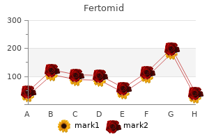
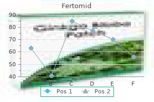
During this period pregnancy x ray risk cheap fertomid amex, keep the vials covered with a clean fine net or muslin cloth to prevent unwanted flies from entering the food vials menstruation vaginal itching cheap fertomid 50 mg with visa. Plug the culture tubes and keep them upright at room temperature in a clean undisturbed area womens health tacoma discount fertomid 50mg visa. Take the culture bottle containing age synchronized wild type Drosophila flies menstrual android discount 50mg fertomid mastercard, tap it gently on the foam pad to let flies fall on the food, quickly remove the cotton plug, taking care not to allow any fly to escape and immediately invert this bottle onto the etherizer (in hour glass arrangement). Care should be taken not to allow the flies touch the medium surface when they are unconscious. Replace the cotton plugs onto the culture vials containing flies, and keep them in horizontal condition until the flies regain consciousness (less than 20 min). Note: Number of flies per tube can be increased or decreased as per requirement Day 3: Look for any dead flies in all the tubes and record the same in the observation book. Introduce flies from the different food vials, one by one, into the negative geotaxis tube, using a small funnel to prevent escape of any fly, tap down the geotaxis tube on the foam pad, and plug the open end of the tube. Let the tube stand vertically and immediately start the stopwatch or start noting time in your wrist watch. Make a minimum of three observations for each tube and calculate average % of flies that climb to the 10 cm height within 10 sec. Plot a bar graph for the % Flies Climbed on Y axis and treatment condition on the X axis. Negative geotaxis assay showing the flies climbing up with different efficiency after feeding on food carrying different concentrations of Rotenone (indicated on each tube). Discard the food vials and other tubes/materials that may carry Rotenone in the labeled waste bin for appropriate disposal. Results/Observations: Calculate the Mean % Flies Climbed under different treatment conditions and record the data in a tabular form as suggested in Table 1. Design an experimental screen to test various drugs/chemicals that may improve climbing activity of Rotenone treated flies for determining their therapeutic value in neurodegenerative disorders. Suppose in your above test, one of the test compounds improves the climbing activity of Rotenone exposed adult flies. A simple and reproducible assay for mobility defects in model organisms such as Drosophila is useful for screening a large number of genotypes or drugs affecting locomotion of flies in a short period of time. Taking advantage of this stereotyped behaviour, it is possible to assess mobility defects. Many studies have characterized degeneration of the dopaminergic neurons by taking advantage of the negative geotaxis behaviour of Drosophila (Chen and Feany, 2005). This modification helps in quantification of mobility defects with an enhanced sensitivity, which helps in detection of mobility phenotype during early stages of disease onset. Flies: Wild type and/or mutant Drosophila flies In: Experiments with Drosophila for Biology Courses. Collect bachelor male and female flies (see Chapter 1) and age for 4-5 days on normal food. Place twenty-five flies in an empty food vial carrying a filter paper soaked in 250 L of 10 mM Paraquat in 5% sucrose solution for 24 or 48 Hr (Phom et al. Watch for next 12 sec and using the graduation marks on the pipette wall, record the distance each fly climbs up in 12 sec. Repeat step 5 and 6 three times for each fly and determine the mean distance climbed up in 12 sec. It should be noted that the climbing assay provides only a primary screen for detection of degeneration of dopaminergic neurons. Further validation by other more specific methods to confirm such neurodegeneration is necessary. Why should defective functioning/degeneration of dopaminergic neurons lead to the mobility defects Does degeneration of neurons other than the dopaminergic ones also lead to mobility defects What other factors may cause the observed mobility defects in the experimental flies Can there be other behavioural phenotypes that can be used as markers for degeneration of dopaminergic neurons
Purchase fertomid 50 mg. The Department of Neurology at Medical Associates.