
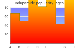
Indapamide
"Indapamide 1.5 mg fast delivery, arterial narrowing".
By: L. Stejnar, M.A., M.D., M.P.H.
Vice Chair, Ponce School of Medicine
By inhibiting cyclooxygenase pulse pressure 2013 effective indapamide 2.5mg, aspirin inhibits the production of prostaglandins and lowers the hypothalamic set-point temperature to its original value arrhythmia generator discount 2.5 mg indapamide. After aspirin treatment arrhythmia or dysrhythmia buy cheap indapamide line, the hypothalamus "reads" the body temperature as being higher than the set-point temperature and activates heat-loss mechanisms arteria labialis superior cheap indapamide online, including sweating and vasodilation of skin blood vessels. The blood values are consistent with acute respiratory alkalosis from hysterical hyperventilation. The tingling and numbness are symptoms of a reduction in serum ionized [Ca2+] that occurs secondary to alkalosis. Because of the reduction in [H+], fewer H+ ions will bind to negatively charged sites on plasma proteins, and more Ca2+ binds (decreasing the free ionized [Ca2+]). Testosterone is converted to its active form, dihydrotestosterone, in some target tissues by the action of 5-reductase. A decrease in radius causes an increase in resistance, as described by the Poiseuille relationship (resistance is inversely proportional to r4). Thus, if radius decreases twofold, the resistance will increase by (2)4, or 16-fold. When heart rate increases, the time between ventricular contractions (for refilling of the ventricles with blood) decreases. Because most ventricular filling occurs during the "reduced" phase, this phase is the most compromised by an increase in heart rate. The blood loss that occurred in the accident caused a decrease in arterial blood pressure. The decrease in arterial pressure was detected by the baroreceptors in the carotid sinus and caused a decrease in the firing rate of the carotid sinus nerves. As a result of the baroreceptor response, sympathetic outflow to the heart and blood vessels increased, and parasympathetic outflow to the heart decreased. A history of head injury with production of dilute urine accompanied by elevated serum osmolarity suggests central diabetes insipidus. Spironolactone inhibits distal tubule Na+ reabsorption and K+ secretion by acting as an aldosterone antagonist. The action potential shown is characteristic of ventricular muscle, with a stable resting membrane potential and a long plateau phase of almost 300 msec. Smooth muscle action potentials would be superimposed on fluctuating baseline potentials (slow waves). Atrial muscle cells of the heart have a much shorter plateau phase and a much shorter overall duration. Depolarization, as in phase 0, is caused by an inward current (defined as the movement of positive charge into the cell). The inward current during phase 0 of the ventricular muscle action potential is caused by opening of Na+ channels in the ventricular muscle cell membrane, movement of Na+ into the cell, and depolarization of the membrane potential toward the Na+ equilibrium potential (approximately +65 mV). Because the plateau phase is a period of stable membrane potential, by definition, the inward and outward currents are equal and balance each other. Phase 2 is the result of opening of Ca2+ channels and inward, not outward, Ca2+ current. In this phase, the cells are refractory to the initiation of another action potential. Phase 2 corresponds to the absolute refractory period, rather than the effective refractory period (which is longer than the plateau). As heart rate increases, the duration of the ventricular action potential decreases, primarily by decreasing the duration of phase 2. The action potential shown represents both depolarization and repolarization of a ventricular muscle cell. The oxidation of I- to I2 is catalyzed by peroxidase and inhibited by propylthiouracil, which can be used in the treatment of hyperthyroidism. The blood values are consistent with metabolic acidosis, as would occur in diabetic ketoacidosis. The subunits of the insulin receptor have tyrosine kinase activity and, when activated by insulin, the receptors autophosphorylate. The phosphorylated receptors then phosphorylate intracellular proteins; this process ultimately results in the physiologic actions of insulin. Blood flow through the artery is proportional to the pressure difference and inversely proportional to the resistance (Q = P/R). Because resistance increased 16-fold when the radius decreased twofold, blood flow must decrease 16-fold.
Systemic vasodilators such as naftidrofuryl oxalate blood pressure chart low bp purchase 2.5 mg indapamide with mastercard, nicotinic acid and thymoxamine (moxisylyte) are also worth trying arteria hepatica propia indapamide 2.5mg fast delivery. Glycerol trinitrate ointment pulse pressure 30 order indapamide with american express, applied once daily may reduce the severity and frequency of attacks and may allow reduction in the dosage of calcium channel blockers and vasodilators hypertension 38 weeks pregnant discount indapamide 2.5 mg free shipping. Infusions with reserpine or prostacyclin help some severe cases although occasionally sympathectomy is needed. Temporal arteritis Here the brunt is borne by the larger vessels of the head and neck. The condition affects elderly people and may be associated with polymyalgia rheumatica. Blindness may follow if the ophthalmic arteries are involved, and to reduce this risk systemic steroids should be given as soon as the diagnosis has been made. Atherosclerosis this occlusive disease, most common in developed countries, will not be discussed in detail here, but involvement of the large arteries of the legs is of concern to dermatologists. These may develop slowly over the years, or within minutes if a thrombus forms on an atheromatous plaque. The feet are cold and pale, the skin is often atrophic, with little hair, and peripheral pulses are diminished or absent. Fasting plasma lipids (cholesterol, triglycerides and lipoproteins) should be checked in the young, especially if there is a family history of vascular disease. Doppler ultrasound measurements help to distinguish atherosclerotic from venous leg ulcers in the elderly (p. Complete assessment is best carried out by a specialist in peripheral vascular disease or a vascular surgeon. Clinical features the sore begins as an area of erythema which progresses to a superficial blister or erosion. If pressure continues, deeper damage occurs with the development of a black eschar which, when removed or shed, reveals a deep ulcer, often colonized by Pseudomonas aeruginosa. The skin overlying the sacrum, greater trochanter, ischial tuberosity, the heel and the lateral malleolus is especially at risk. Arterial emboli Emboli may lodge in arteries supplying the skin and cause gangrene, ulcers or necrotic papules, depending on the size of the vessel obstructed. Causes include dislodged thrombi (usually from areas of atherosclerosis), fat emboli (after major trauma), infected emboli. These are common in patients over 70 years old who are confined to hospital, especially those with a fractured neck of femur. Suitable investigations include venography, Doppler ultrasonography, which can only detect thrombi in large veins at, or above, the popliteal fossa, and 125Ifibrinogen isotope leg scanning. Deep vein thrombosis after a surgical operation is less frequent now, with early postoperative mobilization, regular leg exercises, the use of elastic stockings over the operative period and prophylaxis with low dose heparin. If the affected vein is varicose or superficial it will be red and feel like a tender cord. Migratory superficial thrombophlebitis should arouse suspicion of an underlying malignancy or pancreatic disease. Abnormalities of the vein wall Trauma (operations and injuries) Chemicals (intravenous infusions) Neighbouring infection. This persisting venous hypertension enlarges the capillary bed; white cells accumulate here and are then activated (by hypoxic endothelial cells), releasing oxygen free radicals and other toxic products which cause local tissue destruction and ulceration. The increased venous pressure also forces fibrinogen and 2-macroglobulin out through the capillary walls; these macromolecules trap growth and repair factors so that minor traumatic wounds cannot be repaired and an ulcer develops. Patients with these changes develop lipodermatosclerosis (see below) and have a high serum fibrinogen and reduced blood fibrinolytic activity. Cause Satisfactory venous drainage of the leg requires three sets of veins: deep veins surrounded by muscles; superficial veins; and the veins connecting these togetherathe perforating or communicating veins (Fig. When the leg muscles contract, blood in the deep veins is squeezed back, against gravity, to the heart (the calf muscle pump); reflux is prevented by valves. When the muscles relax, with the help of gravity, blood from the superficial veins passes into the deep veins via the communicating vessels. If the valves in the deep and communicating veins are incompetent, the calf muscle pump now pushes blood into the superficial veins, where the pressure remains high (`venous Venous hypertension is heralded by a feeling of heaviness in the legs and by pitting oedema.
Inform the patient that the test is used to evaluate thrombotic disorders and monitor thrombolytic therapy blood pressure chart for geriatrics buy indapamide with amex. Platelet alloantibodies develop in patients who become sensitized to platelet antigens of transfused blood pulse pressure change with exercise purchase indapamide on line. As a result arrhythmia omega 3 fatty acids buy generic indapamide 2.5mg line, destruction of both donor and native platelets occurs along with a shortened survival time of platelets in the transfusion recipient prehypertension 38 weeks pregnant generic indapamide 2.5mg fast delivery. The platelet antibody detection test is also used for platelet typing, which allows compatible platelets to be transfused to patients with disorders such as aplastic anemia and cancer. Platelet typing decreases the alloimmunization risk resulting from repeated transfusions from random donors. Platelet typing may also provide additional support for a diagnosis of post-transfusional purpura. Inform the patient who has developed platelet antibodies of the importance of taking precautions against bruising and bleeding, including the use of a soft bristle toothbrush, use of an electric razor, avoidance of constipation, avoidance of acetylsalicylic acid and similar products, and avoidance of intramuscular injections. Arterial plethysmography assesses arterial circulation in an upper or lower limb; it is used to diagnose extremity arteriosclerotic disease and to rule out occlusive disease. The test is performed by applying a series of three blood pressure cuffs to the extremity. Venous plethysmography, done with a series of cuffs, measures changes in venous capacity and outflow (volume and rate of outflow); it is used to diagnose a thrombotic condition that causes obstruction of the major veins of the extremity. When the cuffs are applied to an extremity in patients with venous obstruction, no initial increase in leg volume is recorded because the venous volume of the leg cannot dissipate quickly. Body plethysmography measures the total amount (volume) of air within the thorax, whether or not the air is in ventilatory communication with the lung; the elasticity (compliance) of the lungs; and the resistance to airflow in the respiratory tree. It is used in conjunction with pulmonary stress testing and pulmonary function testing. Inform the patient the test is used to measure changes in blood vessel size or changes in gas volume in the lungs. Explain that there may be some discomfort during insertion of the nasoesophageal catheter if compliance testing is done. Instruct the patient to report any unexpected symptoms that occur during the test. Arterial Plethysmography: Explain to the patient that cuffs are applied to the extremity to measure and compare blood flow. Ask the patient to notify medical personnel if he or she has unexpected symptoms during the test. Apply three blood pressure cuffs to the extremity and attach a pulse volume recorder (plethysmograph), which records the amplitude of each pulse wave. When compared with a normal limb, these measurements determine the presence of arterial occlusive disease. Venous Plethysmography: Explain to the patient that cuffs are applied to the extremity to measure and compare blood flow. Apply two blood pressure cuffs to the extremity, one on the proximal part of the extremity (occlusion cuff) and the other on the distal part of the extremity (recorder cuff). Inflate the recorder cuff to 10 mm Hg, and evaluate the effects of respiration on venous volume: Absence of changes during respirations indicates venous thrombotic occlusion. Inflate the occlusion cuff to 50 mm Hg, and record venous volume on the pulse monitor. Deflate the occlusion cuff after the highest volume is recorded in the recorder cuff. A delay in the return to preocclusion volume indicates venous thrombotic occlusion. Body Plethysmography: Place the patient in a sitting position on a chair in the body box. Explain to the patient that the cuffs are applied to the extremities to measure and compare blood flow. Position a nose clip to prevent breathing through the nose, and connect a mouthpiece to a measuring instrument. At the beginning of the study, instruct the patient to pant rapidly and shallowly, without allowing the glottis to close. For compliance testing, a doublelumen nasoesophageal catheter is inserted, and the bag is inflated with air. Impedance Plethysmography: Explain to the patient that cuffs are applied to the extremity to measure and compare blood flow.
Plasma concentration reaches a peak when the drug amount leaving the blood per unit of time equals that being absorbed prehypertension hypertension stage 1 discount indapamide 1.5 mg fast delivery. Drug entry into hepatic and renal tissue constitutes movement into the organs of elimination pulse pressure of 80 purchase indapamide no prescription. The characteristic phasic time course of drug concentration in plasma represents the sum of the constituent processes of absorption prehypertension education purchase genuine indapamide, distribution arrhythmia alcohol purchase 2.5mg indapamide with visa, and elimination, which overlap in time. When distribution takes place significantly faster than elimination, there is an initial rapid and then a greatly retarded fall in the plasma level, the former being designated the -phase (distribution phase), the latter the -phase (elimination phase). Time course of drug concentration Drug concentration in blood (c) Intravenous Intramuscular Subcutaneous Oral Time (t) B. For instance, if two successive doses are omitted, the plasma level will drop below the therapeutic range and a longer period will be required to regain the desired plasma level. In everyday life, patients will be apt to neglect drug intake at the scheduled time. Apart from poor compliance, the same problem may occur when the total daily dose is divided into three individual doses (tid) and the first dose is taken at breakfast, the second at lunch, and the third at supper. Under this condition, the nocturnal dosing interval will be twice the diurnal one. Consequently, plasma levels during the early morning hours may have fallen far below the desired or, possibly, urgently needed range. Time Course of Drug Plasma Levels During Repeated Dosing (A) When a drug is administered at regular intervals over a prolonged period, the rise and fall of drug concentration in blood will be determined by the relationship between the half-life of elimination and the time interval between doses. If the drug amount administered in each dose has been eliminated before the next dose is applied, repeated intake at constant intervals will result in similar plasma levels. If intake occurs before the preceding dose has been eliminated completely, the next dose will add on to the residual amount still present in the body, i. The shorter the dosing interval relative to the elimination half-life, the larger will be the residual amount of drug to which the next dose is added and the more extensively will the drug accumulate in the body. However, at a given dosing frequency, the drug does not accumulate infinitely and a steady state (Css) or accumulation equilibrium is eventually reached. This is so because the activity of elimination processes is concentration-dependent. The higher the drug concentration rises, the greater is the amount eliminated per unit of time. After several doses, the concentration will have climbed to a level at which the amounts eliminated and taken in per unit of time become equal, i. Within this concentration range, the plasma level will continue to rise (peak) and fall (trough) as dosing is continued at a regular interval. The time needed to reach 90 % of the concentration plateau is about 3 times the t1/2 of elimination. Pharmacokinetics 49 Dosing interval Drug concentration Time Dosing interval Time Accumulation: administered drug is not completely eliminated during interval Steady state: drug intake equals elimination during dosing interval Drug concentration Time A. Time course of drug concentration in blood during regular intake Drug concentration Desired therapeutic level? Here, increasing the initial doses (loading dose) will speed up the attainment of equilibrium, which is subsequently maintained with a lower dose (maintenance dose). Change in Elimination Characteristics During Drug Therapy (B) With any drug taken regularly and accumulating to the desired plasma level, it is important to consider that conditions for biotransformation and excretion do not necessarily remain constant.
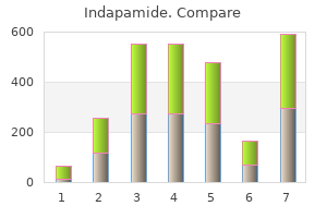
Indapamide 1.5 mg low cost. Fourteen Natural Ways To Lower Your Blood Pressure - Way Cure High Blood Pressure Fast!.